For decades, diagnosing Alzheimer’s disease has remained one of the most frustrating bottlenecks in clinical neurology. By the time an individual begins misplacing everyday items, struggling to find familiar words, or experiencing noticeable lapses in short-term memory, underlying neurodegenerative processes have often been quietly destroying brain tissue for fifteen to twenty years.
Conventional diagnostic pathways rely on invasive lumbar punctures, expensive positron emission tomography (PET) scans, or lengthy neuropsychological examinations. These barriers keep early detection out of reach for the overwhelming majority of the global population, delaying interventions until severe neural damage has already taken place.
Researchers at Arizona State University (ASU), working alongside regional and national medical partners, are pioneering a transformative approach to this challenge: reading the microstructural and vascular changes in the human eye to detect the earliest fingerprints of cognitive decline. By looking directly through the pupil, scientists can inspect living neural tissue non-invasively, creating an affordable, rapid, and universally deployable screening method.
The Eye as an Anatomical Window into the Brain
To understand why ocular diagnostics work for a cerebral disorder, one must examine embryonic development. The retina is not merely an optical sensor connected to the head; it is an embryological extension of the diencephalon, forming directly from neural tube tissue during early fetal growth.
Because the retina shares identical developmental origins with the brain, it contains genuine neurons, glial support structures, and an intricate microvascular network. Crucially, the retina and optic nerve possess an inner blood-retinal barrier that closely mirrors the physiology, selective permeability, and vulnerability of the blood-brain barrier.
When systemic or centralized neurodegenerative cascades occur in the brain, identical biochemical stressors appear inside ocular tissues. Pathological changes do not remain isolated behind the skull; they manifest simultaneously across the fragile cell layers resting at the back of the eye.
Quick Fact: The retina is the only location in the living human body where intact nerves and microscopic blood vessels can be visualized directly, clearly, and without cutting through skin, muscle, or bone.
Because the cornea and lens are naturally transparent, clinicians can illuminate and photograph these neural structures down to single-micron resolutions. This anatomical privilege allows researchers to monitor central nervous system health using non-contact, low-energy optical instruments that take mere seconds to operate.
Pathological Parallels: Amyloid, Tau, and Retinal Microvasculature
Alzheimer’s disease is pathologically characterized by two classical hallmarks: extracellular plaques composed of aggregated amyloid-beta peptides, and intracellular neurofibrillary tangles composed of hyperphosphorylated tau proteins. Historically, tracking these proteins required post-mortem brain tissue autopsies or specialized radiotracer injections.
Pathological investigations have confirmed that amyloid-beta deposits accumulate within the inner nuclear layer and ganglion cell layer of the retina, often preceding measurable neocortical deposition. These ocular plaques mirror the structural dimensions and biochemical properties of cerebral plaques, serving as early peripheral beacons of disease progression.
Simultaneously, hyperphosphorylated tau accumulation within retinal ganglion cells leads to gradual axonal transport failure and structural collapse. This cellular atrophy causes progressive, measurable thinning of the retinal nerve fiber layer (RNFL) and the ganglion cell-inner plexiform layer (GCIPL).
| Pathological Hallmark | Expression in Cerebral Tissue | Expression in Retinal Tissue | Primary Clinical Observation Method |
|---|---|---|---|
| Amyloid-Beta Aggregation | Extracellular senile plaques across cortical and hippocampal areas | Focal deposits in the ganglion cell and inner nuclear layers | Fluorescence imaging and optical reflectivity mapping |
| Hyperphosphorylated Tau | Intracellular neurofibrillary tangles in neurons and synapses | Axonal transport disruption in retinal ganglion cell axons | Cross-sectional optical coherence tomography (OCT) |
| Microvascular Rarefaction | Capillary dropout, reduced cerebral blood flow, vessel tortuosity | Enlarged foveal avascular zone and decreased vessel density | Optical coherence tomography angiography (OCTA) |
| Neuroinflammation | Reactive microgliosis and astrocyte hypertrophy in white/gray matter | Activated retinal microglial migration and perivascular cuffing | High-resolution fundus autofluorescence |
Beyond proteinaceous plaques and tangles, Alzheimer’s disease features a profound vascular component. Cerebral amyloid angiopathy disrupts capillary beds, restricts local perfusion, and triggers hypoxic micro-strokes throughout deep cortical folds.
In the eye, optical coherence tomography angiography reveals identical microvascular breakdown. Patients developing early-stage cognitive decline exhibit significant capillary dropout, abnormal blood vessel branching patterns, and measurable enlargement of the central foveal avascular zone (FAZ).
The ASU Research Approach: Advanced Computational Analysis and Multimodal Scanning
The research programs underway at Arizona State University combine optical instrumentation with sophisticated automated pattern-recognition architectures. A human clinician, no matter how experienced, cannot consistently quantify microscopic retinal thickness differences measured in individual microns or compute fractal dimension changes across millions of capillary pixels.
ASU investigators bridge this gap by deploying computer vision frameworks and statistical machine learning systems trained on multimodal ocular datasets. The system ingests paired data streams from two non-invasive optical capture tools:
- High-Definition Optical Coherence Tomography (OCT): Employs low-coherence near-infrared light waves to capture micrometer-resolution, cross-sectional volumetric slices of retinal tissue layers.
- Optical Coherence Tomography Angiography (OCTA): Tracks the motion of individual red blood cells through successive scans, yielding dynamic, label-free maps of microscopic capillary plexuses without intravenous fluorescent dyes.
Expert Tip: Combining structural cross-sections with functional microvascular flow data dramatically increases diagnostic sensitivity. Relying solely on nerve fiber thinning can produce false positives caused by unrelated conditions like glaucoma or optic neuropathies.
The computational pipeline segments each retinal layer automatically, measuring variations across the macular region and around the optic disc. By calculating the ratio between the ganglion cell layer and surrounding inner retinal bands, the analytical software identifies anomalous structural thinning long before a patient experiences measurable memory decline.
Simultaneously, microvascular algorithms compute fractal dimensions, vessel skeleton density, and lacunarity (the distribution of structural gaps) within the superficial and deep vascular networks. When microvascular rarefaction coincides with localized ganglion cell loss, the system flags the patient as possessing elevated risk profiles for neurodegenerative pathology.
Comparing Detection Paradigms: Eye Scans Versus Traditional Diagnostics
To appreciate the disruptive nature of ocular diagnostics, one must evaluate them against existing clinical standards. Current diagnostic workflows for cognitive decline remain burdensome, painful, and economically prohibitive for routine population-level screening.
The gold standard for observing amyloid accumulation in living patients is Positron Emission Tomography using targeted radiotracers. While exceptionally accurate, amyloid PET scans require cyclotron-produced radioisotopes, specialized nuclear medicine suites, and out-of-pocket patient expenditures that routinely reach thousands of dollars.
Similarly, measuring amyloid-beta and tau ratios through cerebrospinal fluid (CSF) requires a lumbar puncture. This invasive procedure carries risks of post-dural puncture headaches, localized infections, and significant patient apprehension, making it impractical for routine preventive wellness visits.
| Diagnostic Metric | Amyloid PET Brain Scan | Lumbar Puncture (CSF Analysis) | Blood-Based Biomarker Tests | ASU Retinal Optical Screening |
|---|---|---|---|---|
| Invasiveness | Moderate (intravenous radiotracer injection) | High (spinal needle insertion into subarachnoid space) | Low (routine venous blood draw) | Completely non-invasive (non-contact optical scan) |
| Scan / Procedure Time | 45 to 90 minutes | 30 to 60 minutes | 5 to 10 minutes | Under 2 minutes per eye |
| Average Patient Cost | $3,000 – $6,000+ | $1,500 – $3,500 | $300 – $800 | $50 – $150 (projected outpatient fee) |
| Radiation Exposure | Yes (ionizing radiation dose) | None (minor fluoroscopy in complex cases) | None | Completely safe (low-intensity infrared light) |
| Infrastructure Setting | Specialized tertiary hospitals & nuclear labs | Outpatient hospital clinics & surgical suites | Standard phlebotomy laboratories | Optometry clinics, primary care, pharmacy suites |
| Repeatability Frequency | Strictly limited due to radiation and expense | Infrequent due to patient discomfort | Annual or semi-annual monitoring | Unlimited (ideal for quarterly longitudinal tracking) |
While emerging plasma biomarker assays (such as p-tau217 tests) represent a major step forward for decentralized blood testing, they still require centralized clinical laboratories, temperature-controlled sample shipments, and multi-day processing turnarounds. Retinal optical evaluations deliver instantaneous results within the clinical workflow.
The Clinical Workflow: How an Eye-Based Screening Operates
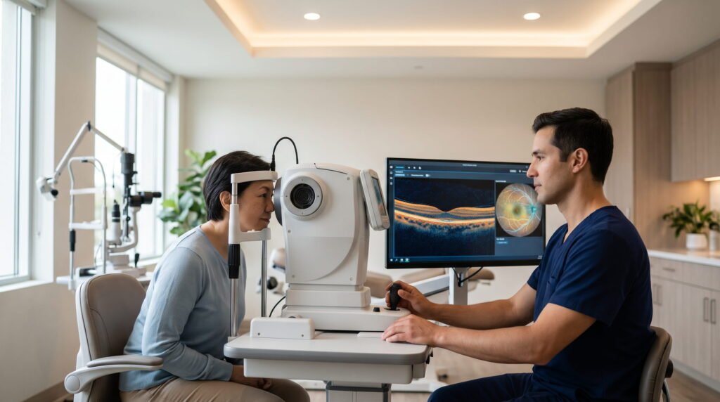
From the patient’s perspective, undergoing an ocular evaluation for neurodegenerative indicators is identical to a standard, non-contact eye examination. The procedure requires no surgical prep, no chemical sedation, and frequently no pharmacological pupil dilation.
The entire diagnostic sequence follows four seamless steps:
- Patient Positioning and Fixation: The patient sits in an ergonomic exam chair and rests their chin and forehead comfortably against cushioned positioning mounts. The individual is instructed to gaze toward a small, dim, colored fixation target inside the instrument aperture.
- Rapid Optical Capture: The technician aligns the optical head with the pupil center. Using safe, invisible near-infrared light, the system records three-dimensional volumetric cubes of the macula and optic nerve head within three to five seconds per eye.
- Automated Feature Extraction: Captured optical slices and flow maps are ingested by the local analysis system. The software identifies internal tissue boundaries, computes layer thicknesses, maps capillary architecture, and strips out image noise or eye-movement artifacts.
- Immediate Clinical Risk Stratification: Within ninety seconds, the software generates a normalized diagnostic report. The report outlines quantitative metrics relative to age-matched normative baselines, categorizing results into low-, moderate-, or high-risk stratifications for cognitive decline.
Pro Tip: Maintaining steady fixation during the brief scanning window ensures optimal image resolution. Modern instruments employ active eye-tracking software that automatically pauses data capture during natural blinks, preventing corrupted data files.
If an anomalous pattern is detected—such as pronounced microvascular dropout coupled with nerve fiber thinning—the primary care physician or optometrist immediately routes the patient to a behavioral neurologist for comprehensive evaluation, cognitive testing, and early therapeutic planning.
Why Early Detection Matters: The Window of Therapeutic Opportunity
A frequent skepticism raised by patients and caregivers asks: “Why bother detecting Alzheimer’s disease twenty years early if there is no absolute cure?” This question misinterprets modern developments in neurotherapeutics and preventive neurology.
Recent pharmaceutical developments have produced monoclonal antibody therapies designed to clear amyloid-beta plaques from cerebral parenchyma. Crucially, phase three clinical trials have shown that these therapies deliver their highest clinical efficacy when administered during the earliest, pre-symptomatic stages of disease, known as Mild Cognitive Impairment (MCI) or subjective cognitive decline.
Once massive neuronal death, cortical atrophy, and synapse destruction have occurred, clearing amyloid plaques cannot restore lost memories or repair destroyed cognitive circuits. The therapeutic goal is preservation, not resurrection.
Important: Identifying neurodegenerative processes during the pre-symptomatic window gives patients a multi-year head start to modify risk factors, enroll in experimental clinical trials, and initiate disease-modifying therapies before functional autonomy is compromised.
Beyond pharmacological agents, early detection empowers individuals to implement aggressive, evidence-backed lifestyle interventions that slow disease progression:
- Cardiovascular Optimization: Strict blood pressure and glycemic management, which preserves cerebral perfusion and prevents vascular dementia overlap.
- Targeted Physical Regimens: Aerobic exercise routines that stimulate Brain-Derived Neurotrophic Factor (BDNF) production and promote adult neurogenesis.
- Sleep Architecture Preservation: Treating obstructive sleep apnea to support optimal glymphatic waste clearance during deep slow-wave sleep cycles.
- Cognitive and Social Enrichment: Building robust cognitive reserve through complex mental tasks, continuous education, and social connectivity.
Technical and Biological Challenges Facing Retinal Biomarkers
Despite the immense promise of ocular diagnostics, transforming research prototypes into everyday diagnostic standards requires solving several formidable technical and biological hurdles. The human eye is not an isolated test tube; it is subject to diverse confounding ocular pathologies that can complicate diagnostic precision.
The most prominent challenge is confounding comorbidity. Conditions such as glaucoma, diabetic retinopathy, macular degeneration, and hypertensive retinopathy alter retinal layer thickness and microvascular morphology in ways that can superficially mimic neurodegenerative changes.
| Confounding Pathological Condition | Overlapping Retinal Manifestation | Distinguishing Diagnostic Criteria Used by ASU Algorithms |
|---|---|---|
| Primary Open-Angle Glaucoma | Progressive thinning of the retinal nerve fiber layer (RNFL) | Glaucoma exhibits localized inferior/superior arcuate defects; Alzheimer’s exhibits generalized diffuse inner macular layer thinning. |
| Diabetic Retinopathy | Capillary non-perfusion, microaneurysms, vascular dropout | Diabetes features characteristic exudates and leakage; neurodegeneration causes vascular rarefaction without blood-retina fluid leakage. |
| Age-Related Macular Degeneration | Focal sub-retinal deposits and outer retinal atrophy | AMD deposits (drusen) settle beneath the retinal pigment epithelium; amyloid deposits reside within the inner retinal layers. |
| Severe Systemic Hypertension | Arteriolar narrowing and microvascular tortuosity changes | Hypertension produces arteriovenous nicking and focal spasms; cognitive decline produces capillary density reduction without mechanical compression. |
Another major obstacle involves equipment variability. Different optical coherence tomography manufacturers utilize varying light wavelengths, scanning speeds, and internal segmentation algorithms. A layer thickness measured on one commercial device may differ by several microns from a measurement taken on a competing brand’s machine.
Common Mistake: Assuming an abnormal retinal thickness reading on an eye scan automatically means a patient has Alzheimer’s disease. Diagnostic algorithms must cross-reference ocular features against normative, age-adjusted baselines and exclude common primary eye diseases before assigning risk.
To overcome these challenges, ASU research teams are developing device-agnostic normalization models. By training algorithms on raw interferometric optical signals rather than post-processed vendor images, researchers ensure diagnostic reliability regardless of which scanning hardware an individual clinic uses.
Ethical, Psychological, and Data Privacy Considerations
The prospect of receiving an early indicator of a neurodegenerative illness during an ordinary optical checkup introduces sensitive ethical and psychological questions. Detecting biomarkers decades before clinical symptom onset alters an individual’s identity, financial planning, and emotional well-being.
Clinicians must address the psychological burden placed on asymptomatic individuals. Learning that one harbors an elevated risk for cognitive decline can induce chronic anxiety, depression, and existential distress if delivered without compassionate counseling, educational context, and concrete management pathways.
Furthermore, robust data governance protocols are essential. High-resolution retinal scans are as unique to an individual as their fingerprints. If retinal diagnostic databases were compromised, an individual’s biometric markers and future health risks could be exposed.
Warning: The legal framework protecting individuals from genetic discrimination (such as the Genetic Information Nondiscrimination Act in the United States) does not universally extend to non-genetic biological markers like retinal optical scans when applying for life insurance, disability insurance, or long-term care policies.
Institutions must maintain strict data sovereignty standards. Patient scans used in research or outpatient screening networks must undergo irreversible anonymization, end-to-end cryptographic encryption, and strict segregation from commercial underwriting ecosystems.
Moreover, patient autonomy must guide clinical deployment. Diagnostic ocular evaluations should operate on an informed-consent model, ensuring that patients retain the right to decide whether they wish to know their pre-symptomatic neurodegenerative status.
The Road to Public Adoption: From Academic Bench to Community Optometry
For this technology to achieve meaningful public health impact, it must transition from elite academic research centers into everyday community healthcare settings. Arizona State University’s strategic vision relies on turning neighborhood optometry and primary care practices into the front lines of neurodegenerative screening.
In the United States alone, tens of millions of adults visit an optometrist or ophthalmologist every year for standard refractive corrections, contact lens fittings, or routine visual acuity checkups. Because modern eye clinics already house commercial optical coherence tomography instruments, the physical hardware is already deployed across thousands of suburban and rural communities.
Integrating neurodegenerative screening does not require clinics to purchase multi-million-dollar imaging devices. Instead, it requires deploying specialized, regulatory-cleared diagnostic software plugins that seamlessly connect to existing optical hardware.
| Implementation Horizon | Primary Focus Objectives | Key Regulatory & Operational Milestones |
|---|---|---|
| Current Phase: Clinical Validation | Multi-site institutional clinical trials; refining segmentation software | Cross-validation against amyloid PET and CSF tau baselines |
| Near-Term (1–3 Years): Regulatory Submission | Formal FDA de novo / 510(k) clearance filings for clinical software | Establishing primary endpoint sensitivity and specificity thresholds |
| Mid-Term (3–5 Years): Clinical Integration | Deployment into regional optometry networks and academic health hubs | Establishing dedicated medical billing codes (CPT) for ocular neural screening |
| Long-Term (5+ Years): Standard of Care | Universal inclusion in annual Medicare and commercial wellness exams | Autonomous edge-processing devices deployed in rural and global mobile clinics |
By establishing dedicated Current Procedural Terminology (CPT) reimbursement codes for preventative ocular neuro-screening, insurers and national healthcare systems can incentivize eye care providers to incorporate these evaluations into annual senior examinations.
This decentralized approach transforms neurodegenerative screening from an intimidating hospital ordeal into an accessible, five-minute component of an annual vision checkup.
Frequently Asked Questions
Can a standard eye exam today tell me if I have Alzheimer’s disease?
No. A routine eye exam performed by an optometrist tests visual acuity, refractive errors, intraocular pressure, and general ocular health. The specialized neuro-retinal algorithms and microvascular analyses developed by ASU are currently operating in research and clinical trial settings and have not yet become standard commercial features across every everyday optical shop.
Does the optical scan hurt, touch the eye, or require dilation drops?
The examination is completely painless, non-contact, and quiet. The patient rests their chin on a support and looks at a small light. Advanced optical coherence tomography instruments capture required data in under ten seconds per eye using harmless infrared light, frequently without requiring pharmacological pupil-dilating eye drops.
How does retinal thinning prove Alzheimer’s rather than normal aging?
While some cellular thinning occurs naturally as humans grow older, Alzheimer’s pathology produces distinct, accelerated, layer-specific atrophy. Specifically, neurodegenerative decline targets the inner retinal layers (the ganglion cell and inner plexiform layers) in specific spatial distributions that deviate measurably from healthy, age-matched normative controls.
Is this technology intended to replace brain PET scans and spinal taps entirely?
Initially, ocular imaging is designed to function as a frontline population screening triage. Patients flagged with high risk by an eye scan can be prioritized for confirmatory blood biomarker tests, PET scans, or lumbar punctures, while individuals with healthy retinal scans can avoid expensive, invasive medical interventions.
Can this technique detect other forms of dementia, such as Parkinson’s or Lewy body dementia?
Yes. Parkinson’s disease, frontotemporal dementia, and Lewy body dementia also manifest distinctive structural and microvascular patterns within the retina. Researchers are currently training distinct mathematical classification profiles to differentiate between the unique retinal signatures associated with these diverse neurological disorders.
What happens if my eye scan shows signs of early cognitive decline?
An anomalous scan is not an immediate life-altering verdict; it is an early warning indicator. If an evaluation reveals elevated risk, your practitioner will connect you with a behavioral neurologist for comprehensive cognitive assessments, blood biomarker verification, lifestyle optimization planning, and consideration of early therapeutic interventions.
How early can these retinal changes be seen before memory loss starts?
Peer-reviewed histological and clinical studies indicate that structural and microvascular changes within the retina can begin ten to twenty years before clinical memory loss, language impairment, or disorientation become outwardly noticeable to friends and family members.
Will health insurance or Medicare cover a retinal scan for Alzheimer’s?
Currently, scans performed specifically for cognitive decline screening are covered primarily within approved clinical research trials. Once these diagnostic software platforms obtain formal regulatory clearance from agencies like the FDA, application for standardized CPT reimbursement codes will enable coverage under Medicare and private health insurance plans.
Are amyloid plaques in the retina identical to those in the cerebral cortex?
Yes. Biochemical and optical analyses have demonstrated that retinal amyloid deposits share the same fundamental beta-sheet protein conformation and immunoreactivity as the classical amyloid plaques found in the hippocampal and cerebral brain tissues of deceased Alzheimer’s patients.
Can eye surgery, such as cataract removal or LASIK, prevent this scan from working?
Standard corneal or lens surgeries (such as LASIK, PRK, or artificial intraocular lens replacements for cataracts) do not prevent optical coherence tomography from capturing the retina. As long as the ocular media remains relatively clear, light can pass through to record accurate cross-sectional data from the back of the eye.
Is the infrared light used during the scan dangerous to the retina?
No. Optical coherence tomography uses low-intensity near-infrared illumination operating well within international eye safety standards established by the American National Standards Institute (ANSI). The energy levels are far lower than natural outdoor daylight exposure and cause no thermal or photochemical tissue damage.
How can individuals volunteer or participate in ASU clinical imaging studies?
Interested community members can connect with Arizona State University’s clinical research portals, the Biodesign Institute, or regional clinical trial registries associated with the Arizona Alzheimer’s Consortium. These initiatives regularly enroll both healthy older adults and individuals experiencing mild memory changes.
The Future of Decentralized Neuro-Diagnostics
The convergence of non-invasive optical instrumentation and computational analysis being led by Arizona State University represents a fundamental turning point in how human society confronts neurodegenerative disease. For more than a century, medicine has treated Alzheimer’s as an intractable mystery locked within the vault of the human skull, diagnosable only after severe cognitive damage had become permanent.
By demonstrating that the back of the human eye serves as a vivid, readable mirror of cerebral microstructures, researchers are breaking down the historic barriers of cost, pain, and geographic isolation that have kept modern diagnostics out of reach. A two-minute optical checkup at a local community vision center has the power to identify microscopic cellular changes decades before a patient forgets a loved one’s name.
The goal of this research is not simply to deliver an earlier diagnosis, but to open a wide, previously inaccessible window of therapeutic opportunity. Armed with early knowledge, patients, families, and physicians can take proactive control of brain health—deploying emerging medical therapies, safeguarding vascular vitality, and preserving the memories and relationships that define a human life.